Registered trialPhase 2
NCT07437547
ClinicalTrials.gov record of NCT07437547 (opens in a new tab)
- Design
- Randomized, double-blind, placebo-controlled
- Enrollment
- 120 (estimated)
- Registry status
- Recruiting
No results posted
Research profile
Pentadecapeptide BPC 157
Research topics, not use categories.
Registered trials2, listed apart from the publications
Content reviewed October 6, 2026
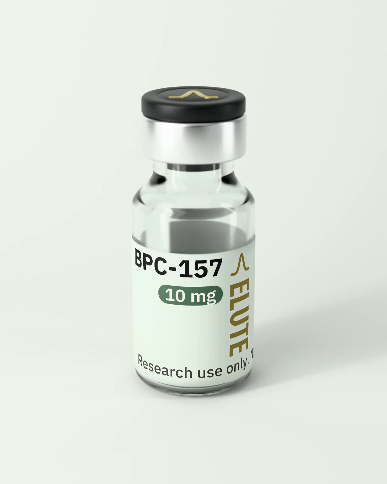
View material (BPC-157 product page)BPC-157 is a short synthetic peptide: a chain of 15 amino acids, the building blocks of proteins. A research group in Zagreb, Croatia, described it in the early 1990s as a fragment of a larger protein that the group had isolated from gastric juice.
Researchers have mainly studied it in rats and in cells grown in the laboratory (cell culture). In rats, it was tested in experimental models in which a tissue is damaged on purpose (the stomach lining, the Achilles at the back of the ankle, a thigh muscle, the skin) or blood flow to a limb or to the brain is interrupted, and the tissue's structure, strength or function is then measured. In cell culture, researchers looked at how fibroblasts (the cells that build connective tissue) and the cells lining blood vessels move, survive and switch particular genes and signaling proteins on or off.
The evidence is at an early stage. Most of it comes from animal and cell studies, much of it from the group that first described the peptide. The human reports listed here are three small studies with no comparison group: a review of 16 patients' records, a pilot study in 12 women and a pilot study in 2 adults. No completed randomized controlled trial with published results was found. Two randomized, placebo-controlled trials are registered: one was recruiting at the last registry check, and the other has an unknown status and no posted results.
The most important limitation is that a result in rats or in cultured cells does not show what happens in people. No receptor for the peptide (a molecule on or in cells that would recognize it) has been identified, and published human data on how the body handles it and on its long-term safety are very limited.
Selected observations from the publications listed in this profile, which are a curated subset of the literature, not all of it. Each one names the system it was observed in and links its source; a finding in cells or animals is not a result in people.
In fibroblasts isolated from rat Achilles tissue, cell migration and spreading increased with the peptide's concentration, together with greater phosphorylation (activation) of the signaling proteins FAK and paxillin; cell proliferation did not change.
(Chang et al. · 2011 · PMID 21030672 (opens PubMed in a new tab))
Limits: Rat cells in culture; the study identified no receptor for the peptide.
In cultured human umbilical vein endothelial cells (cells that line blood vessels), VEGFR2 expression increased and the receptor was internalized, and the VEGFR2–Akt–eNOS pathway was activated over time; the endocytosis inhibitor dynasore blocked the internalization and suppressed the pathway activation.
(Hsieh et al. · 2017 · PMID 27847966 (opens PubMed in a new tab))
Limits: Cell culture. The same paper measured blood flow and vessel counts in a rat model; those results are not summarized here.
In isolated rat aorta, the peptide relaxed the vessel in a concentration-dependent way; the effect was weaker without the vessel's inner lining (endothelium) and was inhibited by L-NAME or hemoglobin, which block nitric oxide. In cultured endothelial cells, phosphorylation of Src, caveolin-1 and eNOS increased.
(Hsieh et al. · 2020 · PMID 33051481 (opens PubMed in a new tab))
Limits: Isolated tissue and cultured cells; no measurement in a living animal or in people.
In a rat model in which blood flow to the brain was stopped for 20 minutes and then restored, hippocampal expression of genes including Egr1, Akt1, Vegfr2 and Nos3 was higher, and expression of Nos2 and Nfkb lower, than in saline controls.
(Vukojević et al. · 2020 · PMID 32558293 (opens PubMed in a new tab))
Limits: A single study by the group that first described the peptide; not repeated by an independent group.
A 2025 systematic review found 36 eligible studies, 35 of them preclinical; the one human study was a retrospective review of patient records, and no controlled human trial was identified.
(Vasireddi et al. · 2025 · PMID 40756949 (opens PubMed in a new tab))
Human, animal and in vitro evidence are kept apart: where a result was observed decides what it can say. In vitro covers cultured cells and tissue studied outside the body. Reviews are listed under Publications; registered trials are listed below, apart from the publications.
The human reports summarized in this profile are three small uncontrolled studies. The first is a retrospective chart review with a telephone survey of 16 people with knee pain of various causes; it had no control group, outcomes were self-reported, and the abstract does not report imaging or validated function instruments. The second is a single-arm, open-label pilot study in 12 women with interstitial cystitis, assessed with a Global Response Assessment questionnaire. The third is an uncontrolled pilot study in 2 adults who had been exposed to the peptide before, with blood markers and vital signs measured at baseline and after exposure; its result is not summarized in this profile. The three reports share a first author, come from private-clinic settings and were published in an alternative-medicine journal. Other human reports exist but are not summarized here: reviews by the originating group mention early trials from the compound's development period whose detailed results were not located in the indexed literature. A 2025 systematic review (36 studies: 35 preclinical and 1 in humans; search through June 2024) found no human safety data, and the single human study it included was retrospective. No completed randomized controlled trial with published results was identified. Two registered trials are listed separately, by their registry entries: a randomized, placebo-controlled trial that was recruiting at its last registry update, with no results, and an earlier registered study whose registry status is unknown and which has no posted results.
Listed publications at this level
Most of the evidence comes from rat models. The models studied include gastric and duodenal lesion models (restraint stress, cysteamine, ethanol), Achilles tenotomy, surgical detachment of the quadriceps muscle, a cutaneous alkali-burn model, hindlimb ischemia and hippocampal ischemia/reperfusion. The endpoints measured include mucosal lesion counts, biomechanical measures (load to failure, Young's modulus), functional indices, histology, extracellular matrix deposition, blood flow and vessel counts, and performance on memory and coordination tests. The authors report differences from control groups on these endpoints; their direction and size are not summarized in this profile. Most of these abstracts do not state sample sizes, blinding is rarely described, and much of the work comes from the originating group. None of the studies listed here used large animals.
Listed publications at this level
The cell and tissue systems studied include primary rat Achilles-derived fibroblasts, Achilles explants, cultured tenocytes, human umbilical vein endothelial cells (HUVEC), NIH 3T3 fibroblasts and isolated rat aorta. In Achilles-derived fibroblasts, faster outgrowth from explants, greater migration and greater survival under oxidative stress were observed, without a direct increase in proliferation, together with FAK–paxillin activation and increased GHR expression. In cultured tenocytes, the peptide alone did not change growth but counteracted growth inhibition by 4-hydroxynonenal. In HUVEC, greater proliferation, migration and tube formation were described, as well as increased VEGFR2 with Akt–eNOS activation. In isolated rat aorta, nitric oxide-dependent vasodilation was observed. None of the studies listed here identified a specific receptor for the peptide, and none used human tenocytes, human gastric mucosa or primary human neurons.
Listed publications at this level
Registry entries, listed apart from the publications and not counted with them. Each links to its ClinicalTrials.gov record and shows only the registered phase, design, enrollment and status, and where its results stand. Results announced by a sponsor are marked as not peer-reviewed and are not summarized here.
ClinicalTrials.gov record of NCT07437547 (opens in a new tab)
No results posted
ClinicalTrials.gov record of NCT02637284 (opens in a new tab)
No results posted
The technical layer: molecular identity, the mechanisms investigated with the evidence level at which each was observed, and the limits of the evidence. The plain-English summary is under Overview.
BPC-157 is a synthetic pentadecapeptide with the sequence GEPPPGKPADDAGLV, the molecular formula C62H98N16O22 and an approximate molecular mass of 1419 Da. It was described in the early 1990s by Sikirić's group (University of Zagreb) as the 15-amino-acid fragment of a larger peptide (BPC) found in gastric juice, and later papers from the same group call it a "stable gastric pentadecapeptide".
It has been studied mainly in rat models of experimental gastric, musculoskeletal, vascular and nervous-system lesions. Proposed mechanistic hypotheses include modulation of the nitric oxide system (Src–caveolin-1–eNOS), VEGFR2–Akt–eNOS signaling in endothelial cells, the FAK–paxillin pathway and GHR expression in Achilles-derived fibroblasts, and changes in early-response genes (Egr-1, c-Fos, c-Jun).
Most of the evidence comes from rat models and cell cultures, much of it from the originating group and a small number of other laboratories; the human reports summarized in this profile are one retrospective chart review and two uncontrolled pilot studies, and no completed randomized controlled trial with published results was identified. It has not received regulatory approval.
Mechanistic hypotheses and observations as they were studied. The evidence level shows where each one was observed; none has been shown in people.
In isolated rat aorta, concentration-dependent vasodilation was observed that was attenuated without endothelium and inhibited by L-NAME or hemoglobin. In cultured endothelial cells, nitric oxide generation and phosphorylation of Src, caveolin-1 and eNOS increased, the phosphorylation changes were abolished by a Src inhibitor, and caveolin-1/eNOS association was reduced. This is a mechanistic hypothesis from one ex vivo and in vitro study; it has not been evaluated in humans.
In human umbilical vein endothelial cells (HUVEC), increased VEGFR2 expression and internalization and time-dependent activation of the VEGFR2–Akt–eNOS pathway were described, with greater tube formation; the endocytosis inhibitor dynasore blocked the internalization and suppressed the pathway activation. The pathway itself was measured only in these cultured cells: in the rat hindlimb ischemia model of the same study, blood flow, vessel counts and vascular VEGFR2 were measured, not Akt or eNOS activation. Another study, in HUVEC and a rat alkali-burn model, reported changes in ERK1/2 phosphorylation and in c-Fos, c-Jun and Egr-1 expression in the endothelial cells, alongside greater endothelial migration. The relevance of this endothelial signaling in humans has not been studied.
In fibroblasts isolated from the rat Achilles, greater migration, cell spreading and F-actin formation were observed, together with a concentration-dependent increase in FAK and paxillin phosphorylation without changes in total protein. No direct effect on proliferation was observed, although survival under oxidative stress (H2O2) was greater. These findings are limited to primary cultures of rat cells.
A microarray analysis in rat Achilles-derived fibroblasts identified GHR, a class I cytokine-family receptor gene, among the most strongly induced genes after exposure to the peptide, with confirmation at the mRNA and protein levels. Subsequent addition of the GHR ligand was associated with greater proliferation and JAK2 activation. This is an in vitro observation whose physiological relevance has not been established.
In a rat model of hippocampal ischemia/reperfusion (bilateral common carotid clamping), hippocampal expression of Egr1, Akt1, Kras, Src, Foxo, Srf, Vegfr2, Nos3 and Nos1 was higher, and that of Nos2 and Nfkb lower, in the BPC-157 group than in saline controls; the study also assessed neuronal damage and performance on memory and coordination tests. These are exploratory findings from a single study by the originating group; they have not been examined by an independent group or in humans.
Each publication is identified by its PubMed ID or DOI and grouped by evidence level. Each heading is our own description of what was studied. Titles, authors and journals are reproduced exactly as published; when the published wording of one of them falls outside our content rules, it is not shown here, and the record links to PubMed, where it can be read in full.
Lee E, Burgess K2025PMID 40131143 (opens PubMed in a new tab)Full record on PubMed
Blood markers of heart, liver, kidney and thyroid function and vital signs were recorded at baseline and after each exposure, and the participants were asked about adverse events at each visit. With two participants, both previously exposed to the peptide, and no control group or blinding, the study cannot characterize safety; its result is not summarized in this profile.
Measured endpoints
Limitations: Uncontrolled; two participants, both previously exposed to the peptide; no blinding; a single private clinic; published in an alternative-medicine journal; a study of this size cannot characterize safety.
“Effect of BPC-157 on Symptoms in Patients with Interstitial Cystitis: A Pilot Study.”
Lee E, Walker C, Ayadi B2024PMID 39325560 (opens PubMed in a new tab)Full record on PubMed
Symptoms were assessed with the Global Response Assessment questionnaire; the authors report responses from all 12 participants and no dropouts. Because this was an open-label pilot study without a control group or blinding and with a subjective outcome, its design does not allow any change to be attributed to the peptide. The self-rated result is not summarized in this profile.
Measured endpoints
Limitations: Small sample (n = 12); no control group or blinding; subjective self-reported outcome; the study procedure itself may have had an effect; a single private clinic; published in an alternative-medicine journal.
Lee E, Padgett B2021PMID 34324435 (opens PubMed in a new tab)Full record on PubMed
Of 17 people on record, 16 were reached by telephone 6 months to 1 year later and asked about their knee pain. There was no control group or blinding, the outcome was self-reported, and the abstract does not report imaging or validated function or quality-of-life instruments; the design carries a high risk of selection and recall bias and does not allow any change to be attributed to the peptide. The self-reported result is not summarized in this profile.
Measured endpoints
Limitations: Retrospective study without a control group or blinding; small sample (n = 16); outcome self-reported by telephone, with no validated scales reported in the abstract; a single private clinic; published in an alternative-medicine journal.
Japjec M, Horvat Pavlov K, Petrovic A et al.Biomedicines2021PMID 34829776 (opens PubMed in a new tab)Title on PubMedDOI 10.3390/biomedicines9111547 (opens the publisher record in a new tab)
In a rat model of surgical detachment of the quadriceps muscle, the BPC-157 and control groups were compared on defect size, function, muscle atrophy, inflammatory infiltrate and the orientation of the junction tissue from day 7 to day 42, together with eNOS and COX-2 mRNA, oxidative stress and nitric oxide levels; the direction and size of these differences are not summarized in this profile.
Measured endpoints
Limitations: Animal model; no human data; sample size not stated in the abstract; study from the originating group; blinding not described.
Vukojević J, Vrdoljak B, Malekinušić D et al.Brain and Behavior2020PMID 32558293 (opens PubMed in a new tab)Title on PubMedDOI 10.1002/brb3.1726 (opens the publisher record in a new tab)
In rats subjected to cerebral ischemia/reperfusion, hippocampal expression of Egr1, Akt1, Kras, Src, Foxo, Srf, Vegfr2, Nos3 and Nos1 was higher, and that of Nos2 and Nfkb lower, in the BPC-157 group than in saline controls; Mapk1 was not activated. Early and late hippocampal neuronal damage and performance on memory (Morris water maze), locomotion and coordination tests were also assessed; their direction is not summarized in this profile. Exploratory findings in an animal model.
Measured endpoints
Limitations: Animal model; no human data; sample size not stated in the abstract; study from the originating group; blinding not described.
Hsieh MJ, Liu HT, Wang CN et al.Journal of Molecular Medicine (Berlin)2017PMID 27847966 (opens PubMed in a new tab)Title on PubMedDOI 10.1007/s00109-016-1488-y (opens the publisher record in a new tab)
In HUVEC, VEGFR2 mRNA and protein increased (VEGF-A did not), with internalization of the receptor, time-dependent activation of the VEGFR2–Akt–eNOS pathway and greater tube formation; these effects were suppressed by the endocytosis inhibitor dynasore. Vascular density in the chorioallantoic membrane assay, and blood flow (laser Doppler), vessel counts and vascular VEGFR2 in rats with hindlimb ischemia, were also measured; the direction and size of these in vivo endpoints are not summarized in this profile.
Measured endpoints
Limitations: Animal model and cell cultures; no human data; sample size not stated in the abstract.
Huang T, Zhang K, Sun L et al.2015PMID 25995620 (opens PubMed in a new tab)Full record on PubMedDOI 10.2147/DDDT.S82030 (opens the publisher record in a new tab)
In a rat cutaneous alkali-burn model, the BPC-157 and control groups were compared on the size of the burned area, granulation tissue, re-epithelialization, extracellular matrix deposition and tissue VEGF; the direction and size of these endpoints are not summarized in this profile. In HUVEC, greater proliferation, migration and tube formation were described, with regulation of ERK1/2 phosphorylation and of c-Fos, c-Jun and Egr-1 expression; these cell findings refer to HUVEC, not to the NIH 3T3 cells also used. Study by a group independent of the originating group.
Measured endpoints
Limitations: Animal model and cell cultures; no human data; sample size not stated in the abstract.
Staresinic M, Sebecic B, Patrlj L et al.Journal of Orthopaedic Research2003PMID 14554208 (opens PubMed in a new tab)Title on PubMedDOI 10.1016/S0736-0266(03)00110-4 (opens the publisher record in a new tab)
After complete Achilles tenotomy in rats, the BPC-157 and saline groups were compared on biomechanical measures (load to failure, Young's modulus), the Achilles functional index, the size of the macroscopic defect and histology (inflammatory cells, fibroblasts, reticulin and fibrillar extracellular matrix); the direction and size of the differences are not summarized in this profile. In cultured tenocytes, the peptide alone did not change growth but counteracted the growth inhibition caused by the aldehyde 4-hydroxynonenal.
Measured endpoints
Limitations: Animal model; no human data; sample size not stated in the abstract; blinding not described; study from the originating group.
Sikiric P, Seiwerth S, Grabarevic Z et al.Life Sciences1994PMID 7904712 (opens PubMed in a new tab)Title on PubMedDOI 10.1016/0024-3205(94)00796-9 (opens the publisher record in a new tab)
In three rat models of gastric and duodenal lesions, mucosal lesions were counted after BPC-157 and after each of the reference agents, and Monastral blue labeling was used to examine the mucosal endothelium. The authors report differences between BPC-157 and the reference agents; their direction and size are not summarized in this profile. Early primary study in an animal model.
Measured endpoints
Limitations: Animal model; no human data; sample size not stated in the abstract; blinding not described.
“Modulatory effects of BPC 157 on vasomotor tone and the activation of Src-Caveolin-1-endothelial nitric oxide synthase pathway.”
Hsieh MJ, Lee CH, Chueh HY et al.Scientific Reports2020PMID 33051481 (opens PubMed in a new tab)DOI 10.1038/s41598-020-74022-y (opens the publisher record in a new tab)
In isolated rat aorta, concentration-dependent vasodilation was observed that was attenuated in the absence of endothelium and inhibited by L-NAME or hemoglobin, indicating a nitric oxide-dependent mechanism. In endothelial cells, nitric oxide generation and phosphorylation of Src, caveolin-1 and eNOS increased and were abolished by a Src inhibitor, with less caveolin-1/eNOS association in co-immunoprecipitation assays. No direct relaxant effect on smooth muscle cells was seen in the 3D model.
Measured endpoints
Limitations: Ex vivo and in vitro systems; no in vivo animal or human data.
Chang CH, Tsai WC, Hsu YH et al.Molecules2014PMID 25415472 (opens PubMed in a new tab)Title on PubMedDOI 10.3390/molecules191119066 (opens the publisher record in a new tab)
Microarray analysis identified GHR among the most strongly induced genes in Achilles-derived fibroblasts exposed to the peptide; the concentration- and time-dependent increase was confirmed at the mRNA and protein levels. Subsequent stimulation of pre-exposed cells with the GHR ligand was associated with greater proliferation (MTT, PCNA) and JAK2 activation. In vitro observation whose in vivo relevance was not assessed.
Measured endpoints
Limitations: In vitro study in rat cells; no animal or human data; the proliferation finding depended on exogenous addition of the GHR ligand.
Chang CH, Tsai WC, Lin MS et al.Journal of Applied Physiology2011PMID 21030672 (opens PubMed in a new tab)Title on PubMedDOI 10.1152/japplphysiol.00945.2010 (opens the publisher record in a new tab)
In rat Achilles explants, cell outgrowth was faster; fibroblast proliferation (MTT) did not change, but survival under H2O2 stress, transwell migration and cell spreading increased in a concentration-dependent manner, with F-actin formation and higher FAK and paxillin phosphorylation and no change in total protein. The authors propose the FAK–paxillin pathway as a mechanistic hypothesis.
Measured endpoints
Limitations: In vitro study in rat cells; no in vivo animal or human data; no receptor was identified.
“Emerging Use of BPC-157 in Orthopaedic Sports Medicine: A Systematic Review.”
Vasireddi N, Hahamyan H, Salata MJ et al.HSS Journal2025PMID 40756949 (opens PubMed in a new tab)DOI 10.1177/15563316251355551 (opens the publisher record in a new tab)
Of 544 records identified, 36 studies were included (35 preclinical and 1 in humans). The authors synthesize functional, structural and biomechanical measures from preclinical musculoskeletal models, together with GHR expression and inflammatory cytokine levels; the only study in humans was retrospective. No human safety data were found, and the evidence was graded as level IV and V.
Measured endpoints
Limitations: Included evidence graded level IV and V; a single study in humans (retrospective); heterogeneous models; no human safety data were located.
Gwyer D, Wragg NM, Wilson SLCell and Tissue Research2019PMID 30915550 (opens PubMed in a new tab)Title on PubMedDOI 10.1007/s00441-019-03016-8 (opens the publisher record in a new tab)
Critical review by an independent group (Loughborough University) of the preclinical literature in small rodent models of muscle and connective-tissue lesions. It notes that only a handful of research groups had studied the peptide in depth, that the effects reported in rodents had yet to be confirmed in humans and that the precise mechanisms still needed to be understood.
Measured endpoints
Limitations: Narrative review without a systematic methodology; the evidence reviewed comes almost exclusively from animal studies.
Sikirić P, Petek M, Rucman R et al.Journal of Physiology, Paris1993PMID 8298609 (opens PubMed in a new tab)Title on PubMedDOI 10.1016/0928-4257(93)90038-u (opens the publisher record in a new tab)
Early overview by the originating group of its own work. It refers back to the group's isolation of a gastric juice peptide (BPC, Mr about 40,000), presents the fully characterized 15-amino-acid fragment BPC 157, which the authors consider essential for the activity they studied, and sets out their hypothesis that the stomach mediates responses to stress in other organs. It summarizes the group's lesion models in several organs and proposes the peptide as a possible endogenous mediator.
Measured endpoints
Limitations: Narrative review of the authors' own work; no human data; the text itself acknowledges that the ultimate mechanism is poorly understood.
For laboratory research use only. Not for human or veterinary use or consumption. Not for diagnostic use. Not evaluated by the U.S. FDA.